Blog Archives
Anatomy – Nervous System
The Anatomy folders contain approximately seven hundred images and have the following arrangement: Anatomy, Anatomy – Animals, Anatomy – Eyes, Anatomy -Hands, and Anatomy – Nervous System. Like the other Anatomy categories, Anatomy – Nervous System consists of photographs of both the interior and exterior, microscopic views, medical illustrations, and creative (non-technical) illustrations.

© 1947, 1981, 1986 Anatomical Chart Company. Chicago, Illinois. Anatomical Illustration by Peter Bachin.

Dissected Neurons. Life Oct. 22, 1971. “Subtle beauties, baffling complexities.”

[At the Synapse, a sudden chemical invasion] “This painting, which is based on the actual specimen…depicts how a signal gets from one neuron to another. Here an axon (the big horizontal shape) forms two synapses, the first with a dendrite offshoot (vertical trunk on the left) and the other with the dendrite itself (right).” Life. October 22, 1971.
![[Succor and support from the gluey glia] Glia Cells. "Greek word for glue, the sticky glia's chief function seems to be to service neurons." Life. Oct. 22, 1971.](https://svapicsandmags.com/wp-content/uploads/2013/04/glia-life-oct-22-1971.jpg)
[Succor and support from the gluey glia] Glia Cells. “Aptly named for the Greek word for glue, the sticky glia’s chief function seems to be to service neurons.” Life. Oct. 22, 1971.

Man Holding Brain

“The interior view of the base of the skull (seen though a wide-angel lens) shows the openings in the bone for the spinal cord and the two jugular veins (bottom), and the two optic nerves (center). Life. Oct. 1, 1971.

© 1983, 1986 Anatomical Chart Co., Chicago, IL.. Illustrated by Ernest W. Beck, medical illustrator, in consultation with Harry Monsen, Ph.D., Professor of Anatomy, College of Medicine, University of Illinois.
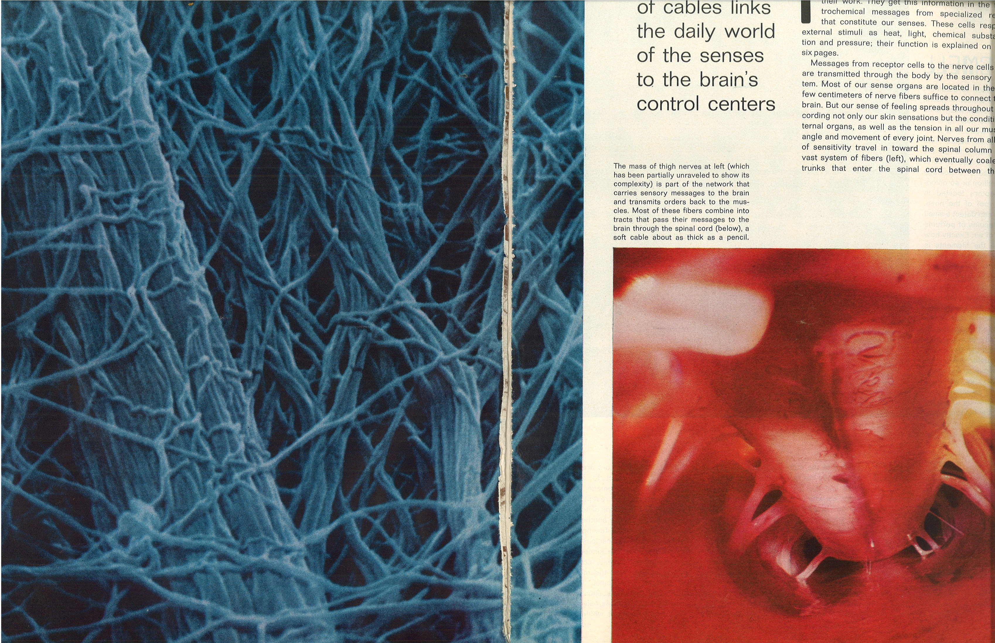
Thigh Nerves (left) Spinal Cord (right). Life Oct. 1, 1971.
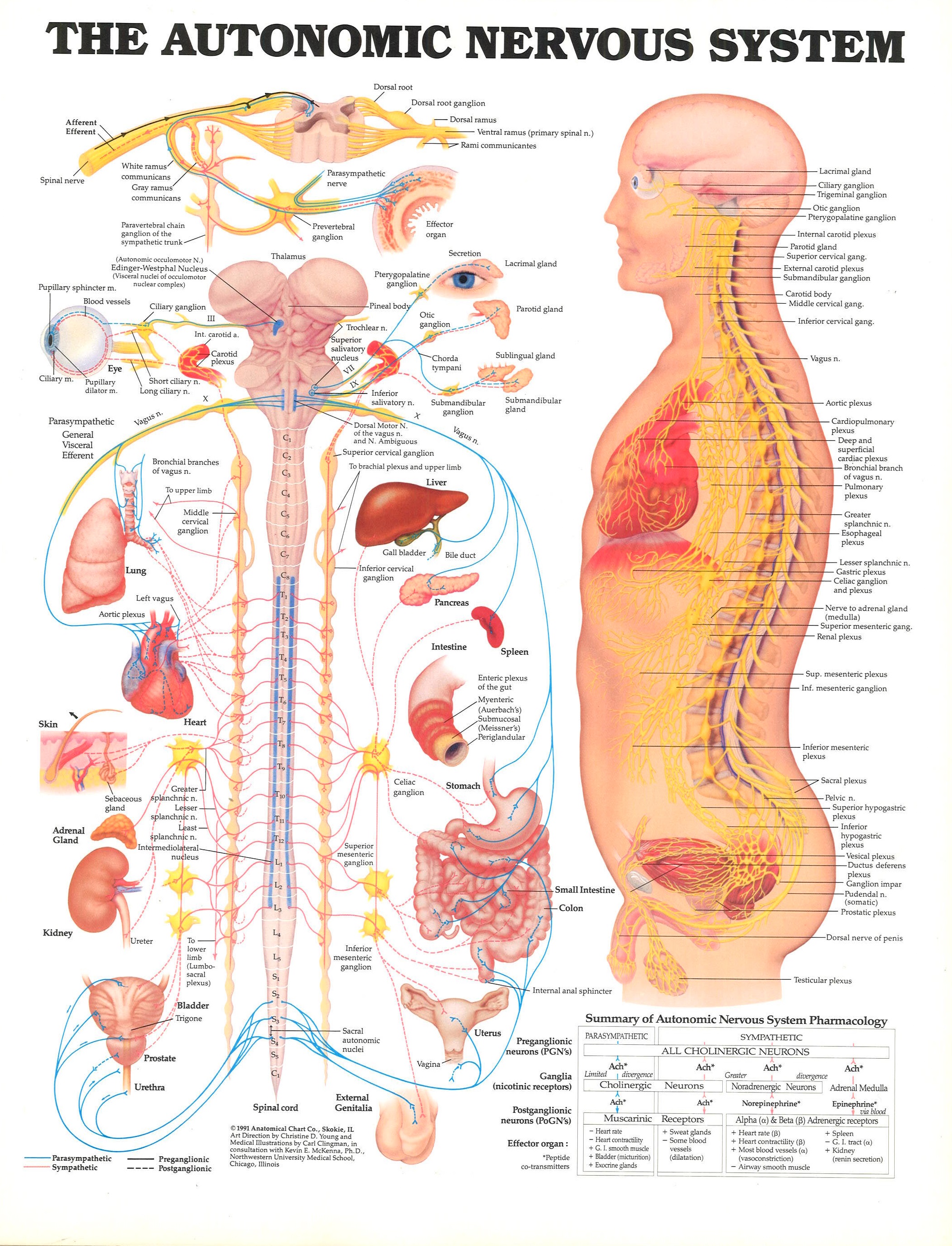
© 1991 Anatomical Chart Co., Skokie, IL. Art Direction by Christine D. Young and Medical Illustrations by Carl Clingman, in consultation with Kevin E. McKenna, Ph.D., Northwestern University Medical School, Chicago, Illinois.

