Category Archives: Picture Collection
People – Sleeping
Here is a sampling of People – Sleeping, a folder containing 60 pictures of bodies whose minds are elsewhere.

© Life. April 24, 1939.


Photos by Michael Putnam.
Left: Patna, 1966.
Right: new York City, 1975.

Photos by Michael Putnam.
New York City.
Left: 1970.
Right: 1975.

Photos by Michael Putnam.
Left: Los Angeles, 1977.
Right: New York City, 1976.
Circus – Clowns
The Circus category has been subdivided. Along with a general folder, you will find Circus – Acrobats, Circus – Clowns, and Circus – Posters & Advertisements. Please enjoy the below selections from the Clowns folder, which has 42 items packed clownishly into one folder. They run the gamut, from the manic and frightening, to the heartbroken and cabbage-coddling.

© National Geographic Society 1948. Photos by Harold E Edgerton and Edwin L Wisherd.


Sunpak Camera Ad

© Geo, Volume II, June 1980. Photo: Richard D Gordon. This clown is “One of the actors in a skit on the Gang of Four.”

© National Geographic Society 1948. Photo by J Baylor Roberts.


Ringling Bros Barnum & Bailey “Circus Clown Alley” Troupe
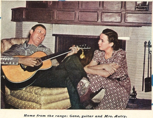
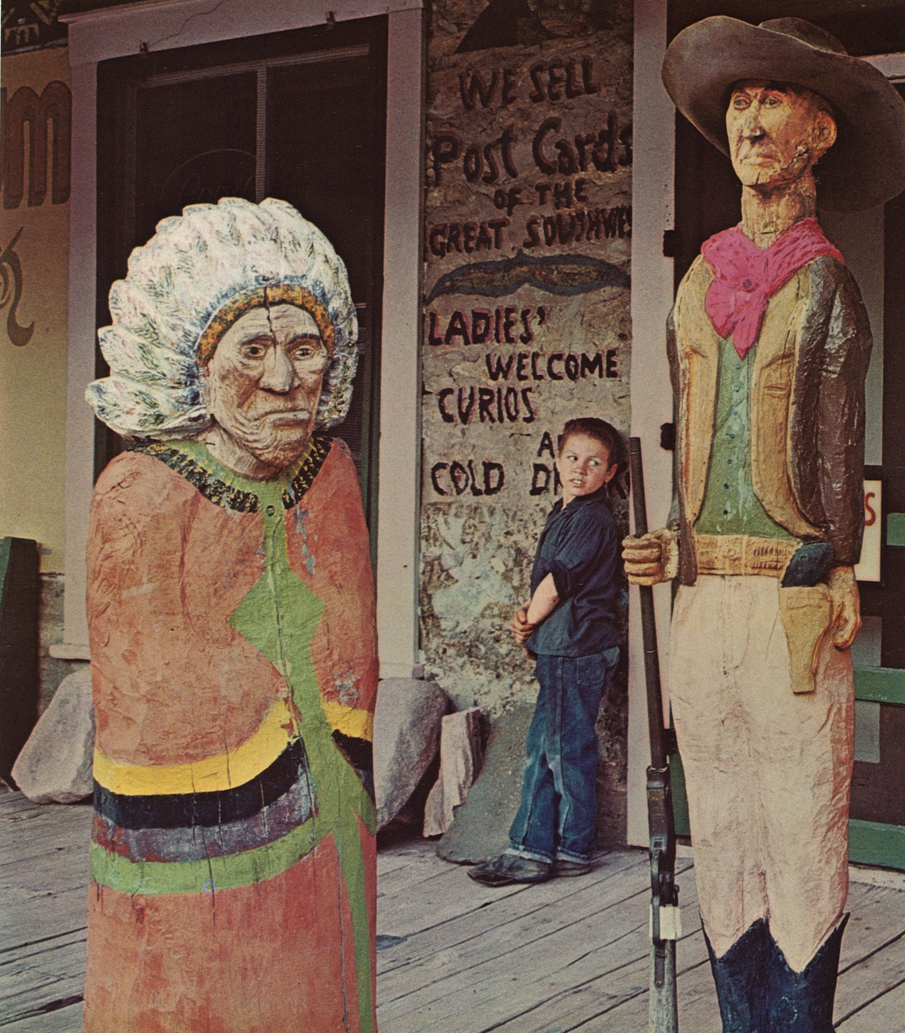
Cowboys & Western Life
These fine as cream gravy ‘curly wolves’ are cuttin’ a swell in apple pie order, and I’m no flannel mouth. Pony up! Quit beatin’ the devil around the stump and get a wiggle on to the collection to see for yourself.
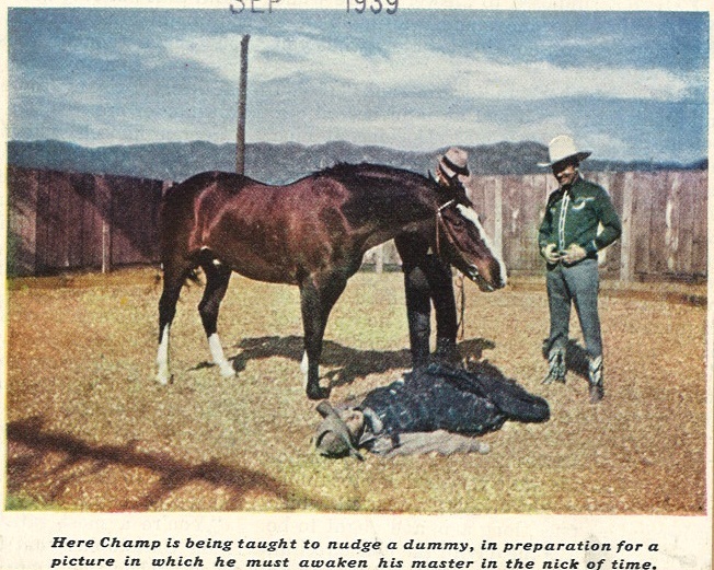
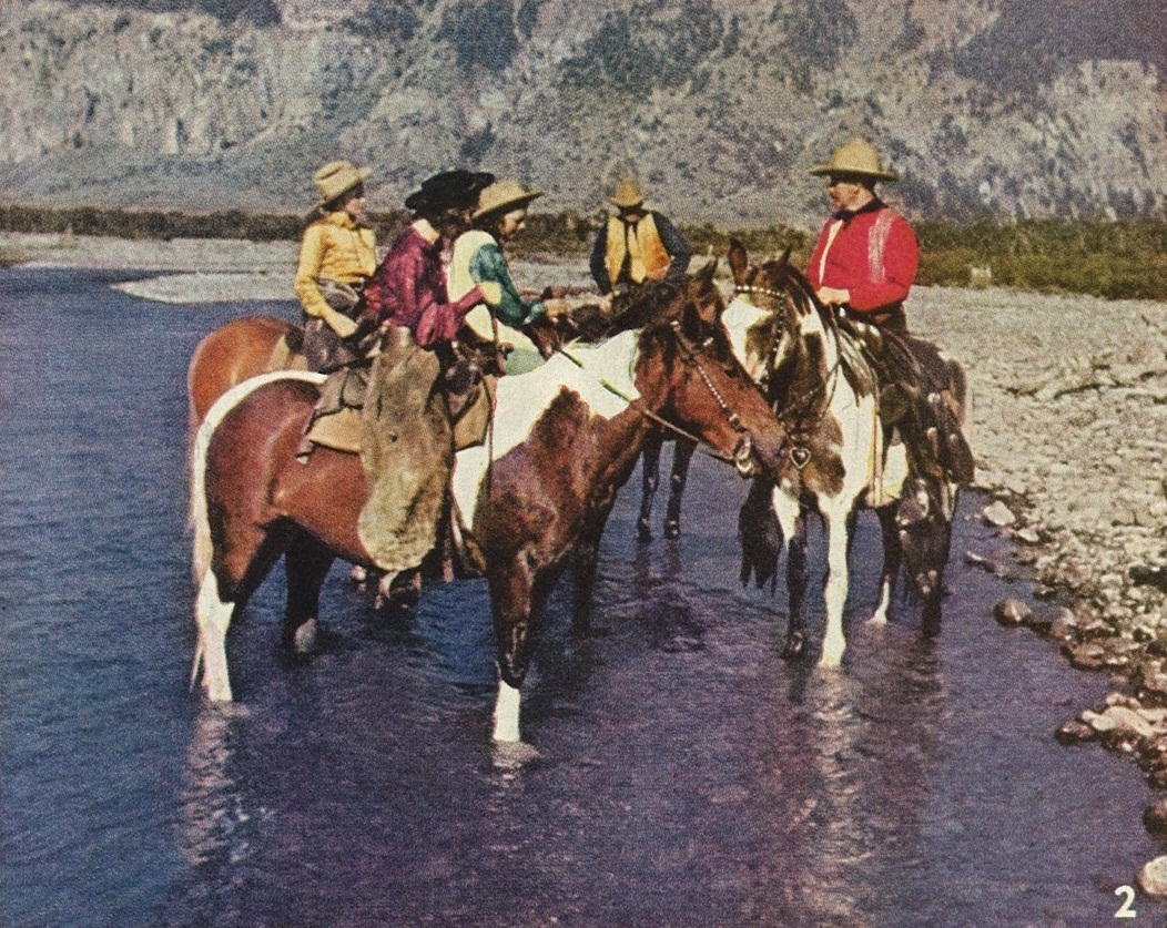
A sample from the 134 items in Cowboys & Western Life follows.


September 1939.
Photo: Ivan Dimitri.
September 1939.

1929.

1929.
September 1939.

September 1939.
June 1939.

Deadwood Dick (Nat Love)

May 1956
America Illustrated, 1967.


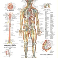
Anatomy – Nervous System
The Anatomy folders contain approximately seven hundred images and have the following arrangement: Anatomy, Anatomy – Animals, Anatomy – Eyes, Anatomy -Hands, and Anatomy – Nervous System. Like the other Anatomy categories, Anatomy – Nervous System consists of photographs of both the interior and exterior, microscopic views, medical illustrations, and creative (non-technical) illustrations.

© 1947, 1981, 1986 Anatomical Chart Company. Chicago, Illinois. Anatomical Illustration by Peter Bachin.

Dissected Neurons. Life Oct. 22, 1971. “Subtle beauties, baffling complexities.”

[At the Synapse, a sudden chemical invasion] “This painting, which is based on the actual specimen…depicts how a signal gets from one neuron to another. Here an axon (the big horizontal shape) forms two synapses, the first with a dendrite offshoot (vertical trunk on the left) and the other with the dendrite itself (right).” Life. October 22, 1971.
![[Succor and support from the gluey glia] Glia Cells. "Greek word for glue, the sticky glia's chief function seems to be to service neurons." Life. Oct. 22, 1971.](https://svapicsandmags.com/wp-content/uploads/2013/04/glia-life-oct-22-1971.jpg)
[Succor and support from the gluey glia] Glia Cells. “Aptly named for the Greek word for glue, the sticky glia’s chief function seems to be to service neurons.” Life. Oct. 22, 1971.

Man Holding Brain

“The interior view of the base of the skull (seen though a wide-angel lens) shows the openings in the bone for the spinal cord and the two jugular veins (bottom), and the two optic nerves (center). Life. Oct. 1, 1971.

© 1983, 1986 Anatomical Chart Co., Chicago, IL.. Illustrated by Ernest W. Beck, medical illustrator, in consultation with Harry Monsen, Ph.D., Professor of Anatomy, College of Medicine, University of Illinois.
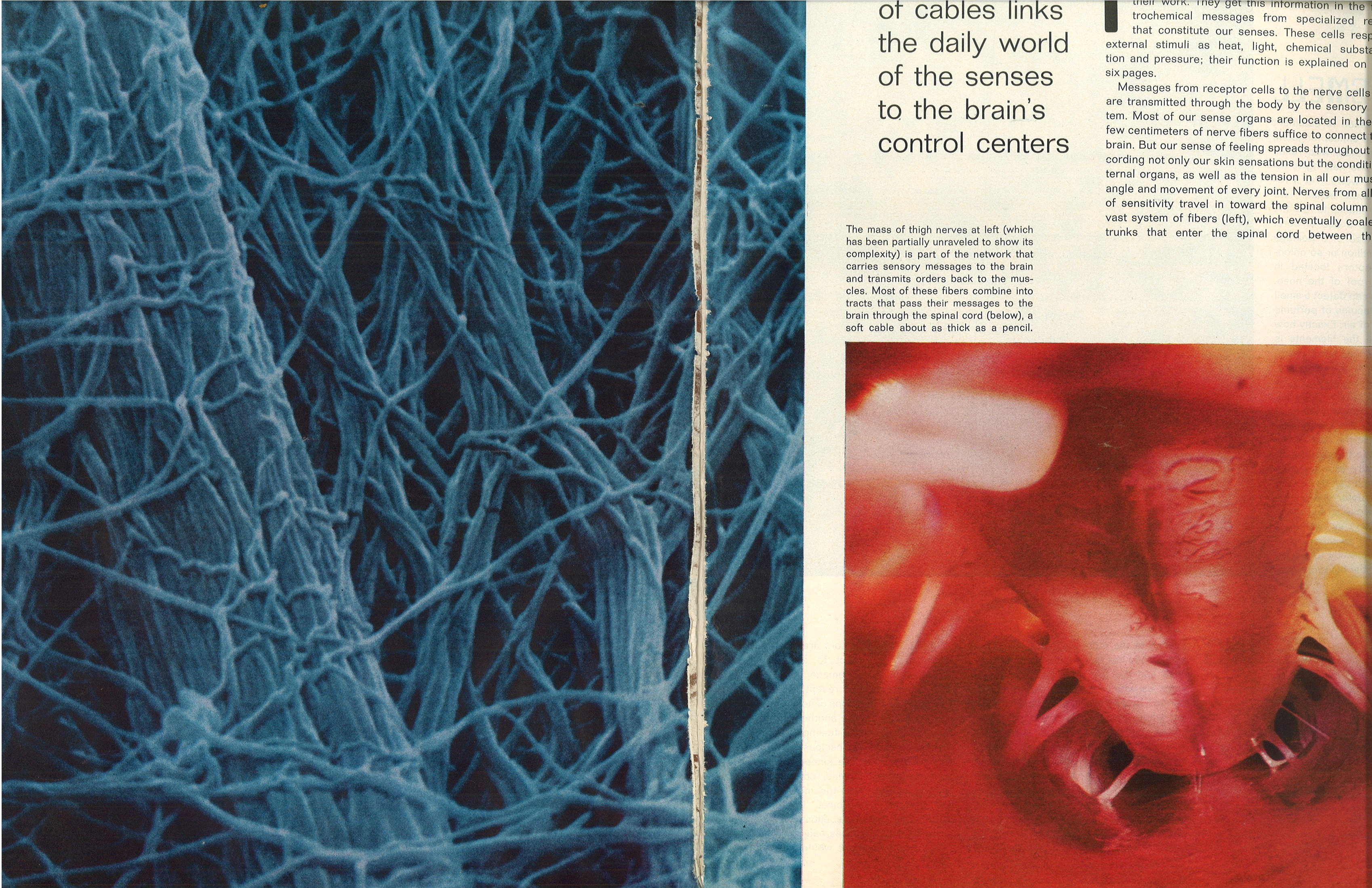
Thigh Nerves (left) Spinal Cord (right). Life Oct. 1, 1971.
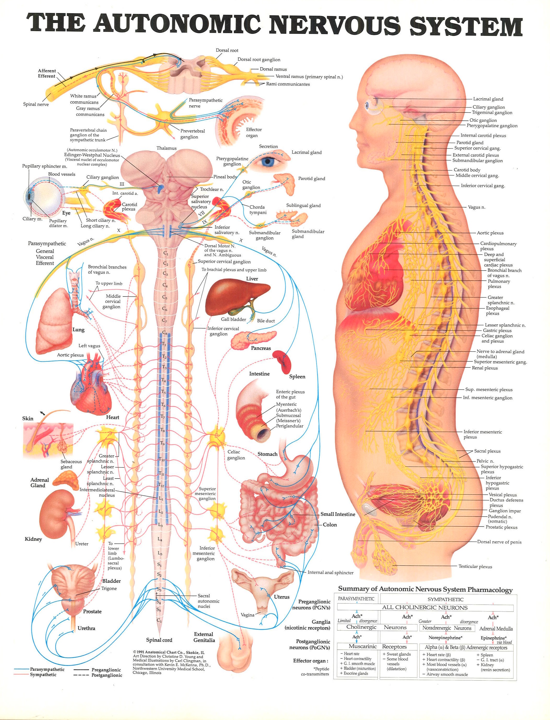
© 1991 Anatomical Chart Co., Skokie, IL. Art Direction by Christine D. Young and Medical Illustrations by Carl Clingman, in consultation with Kevin E. McKenna, Ph.D., Northwestern University Medical School, Chicago, Illinois.
Animals – Assorted
Spread throughout three folders are the subcategories Animals – Assorted (A-G), (H-N), and (O-Z). These folders are home to about 70 images of animals that do not fall into any of the Picture Collection’s current animal subcategories (there are over 60 subcategories of Animals with thousand of pictures, from Apes and Monkeys to Zebras). Maybe if some of these adorable little creatures find a few more like themselves, they too will have the distinction of having their own folder. I would love to see a folder full of proud platypuses.

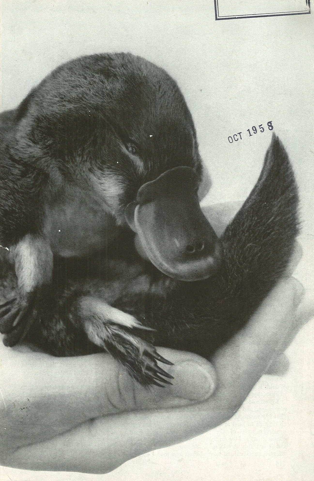
Platypus

Sloth
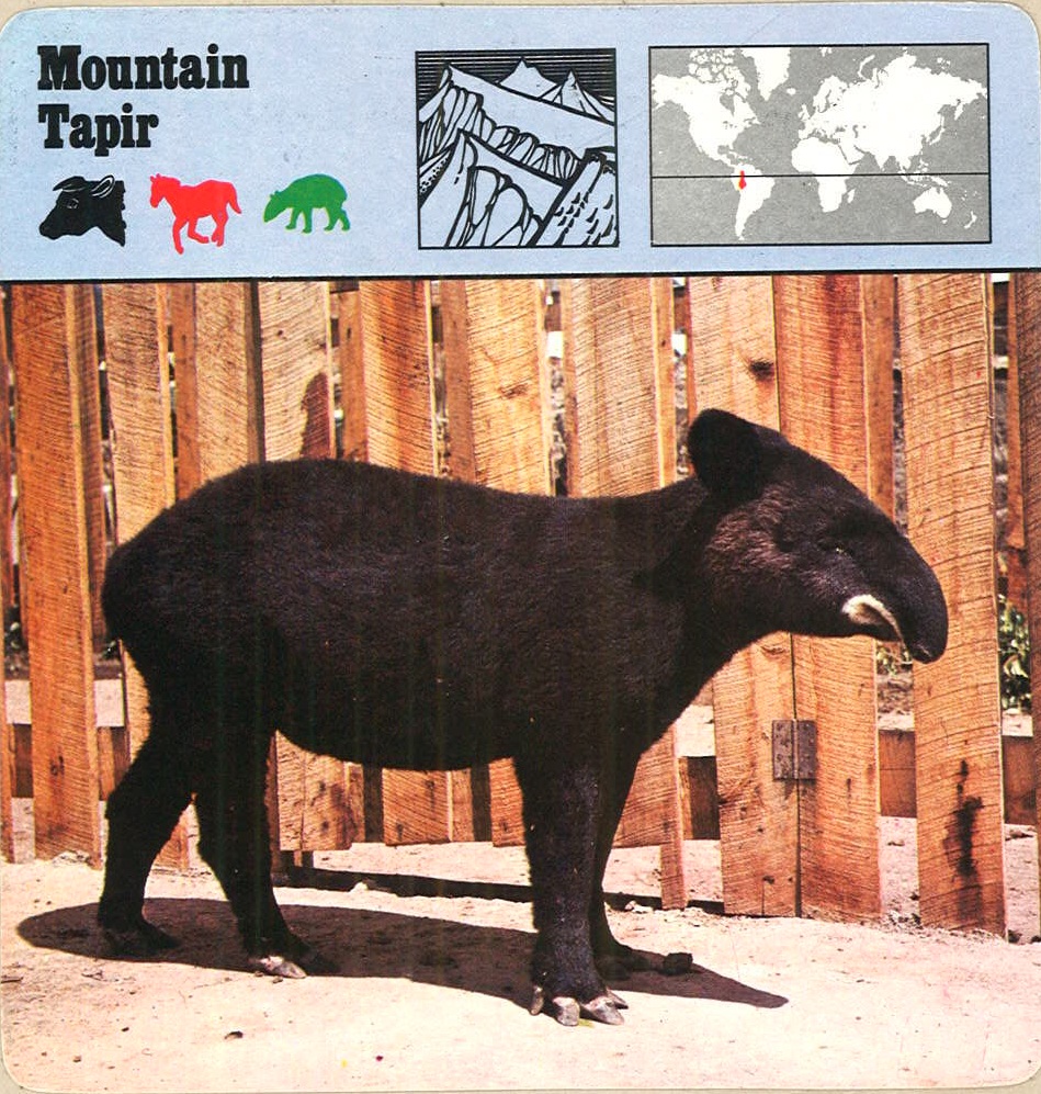
Mountain Tapir

Galagos, commonly known as ‘Bush Babies’ and also called ‘Nagapies’–meaning “Little night monkeys” in Afrikaans

Making a rare appearance above ground, a common European mole prepares to burrow its way back into the security of a subsurface tunnel, using paddle-like appendages to scrape soil and push it rearward or up.
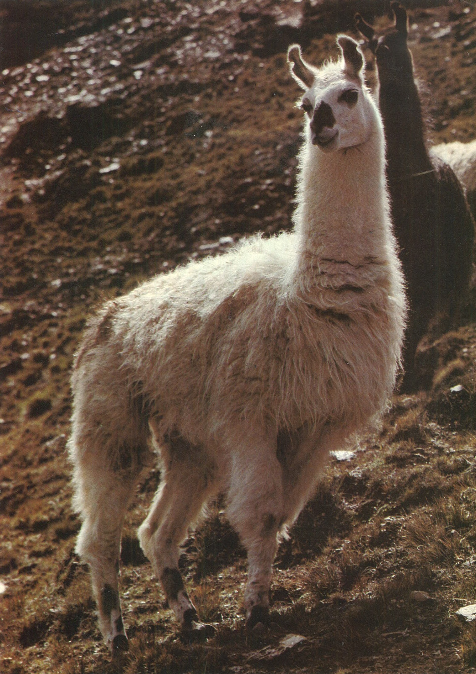
Llama
National Geographic, Volume 219, Issue Number 05, May 2011.
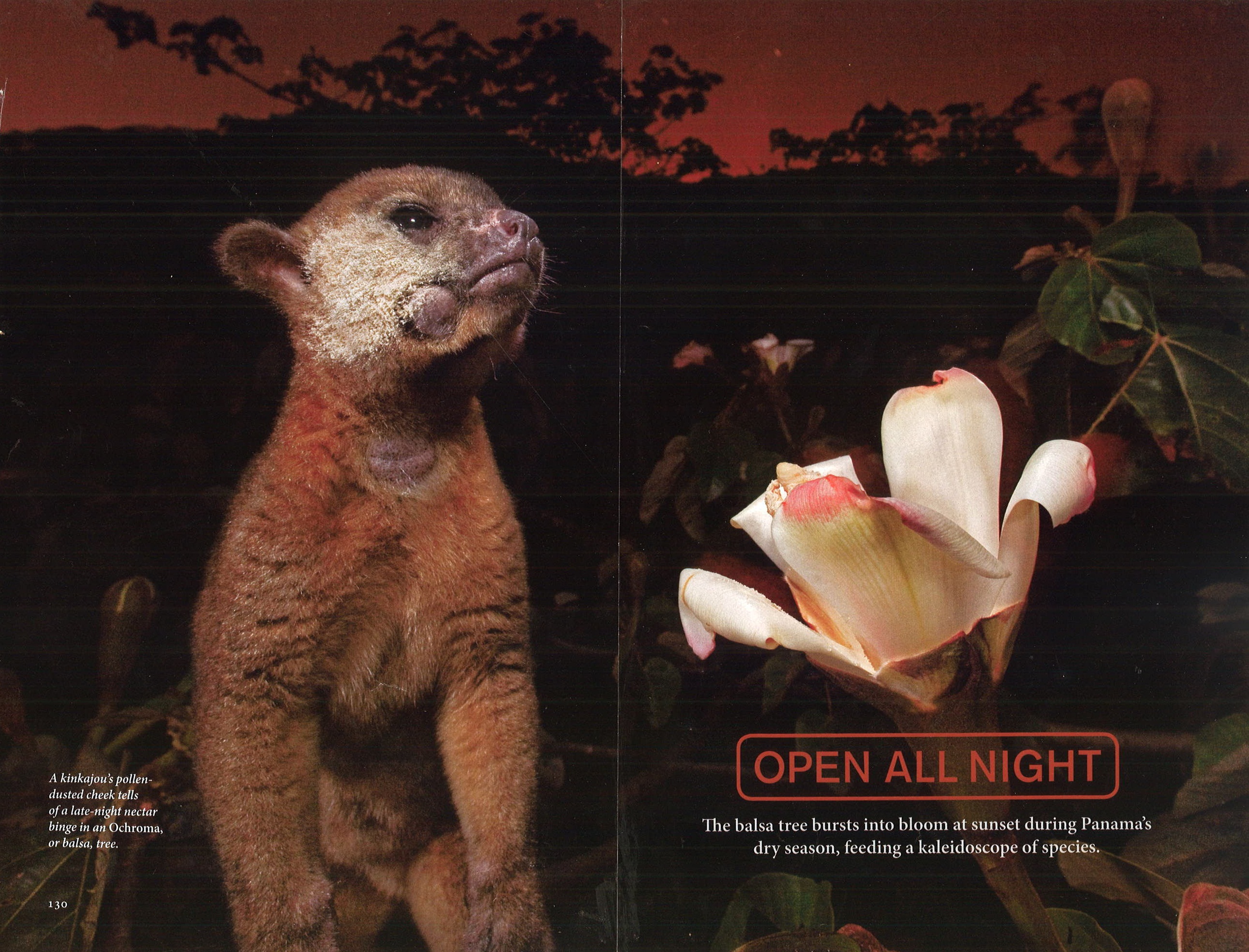
A Kinkajou’s pollen-dusted cheek tells of a late-night nectar binge in an Ochroma, or balsa, tree.

Aardvark
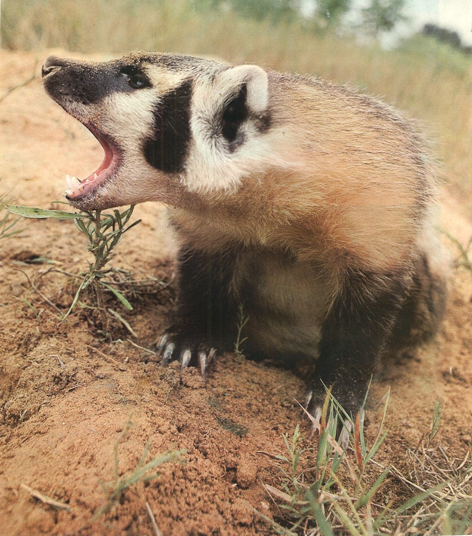
Badger

Anteater
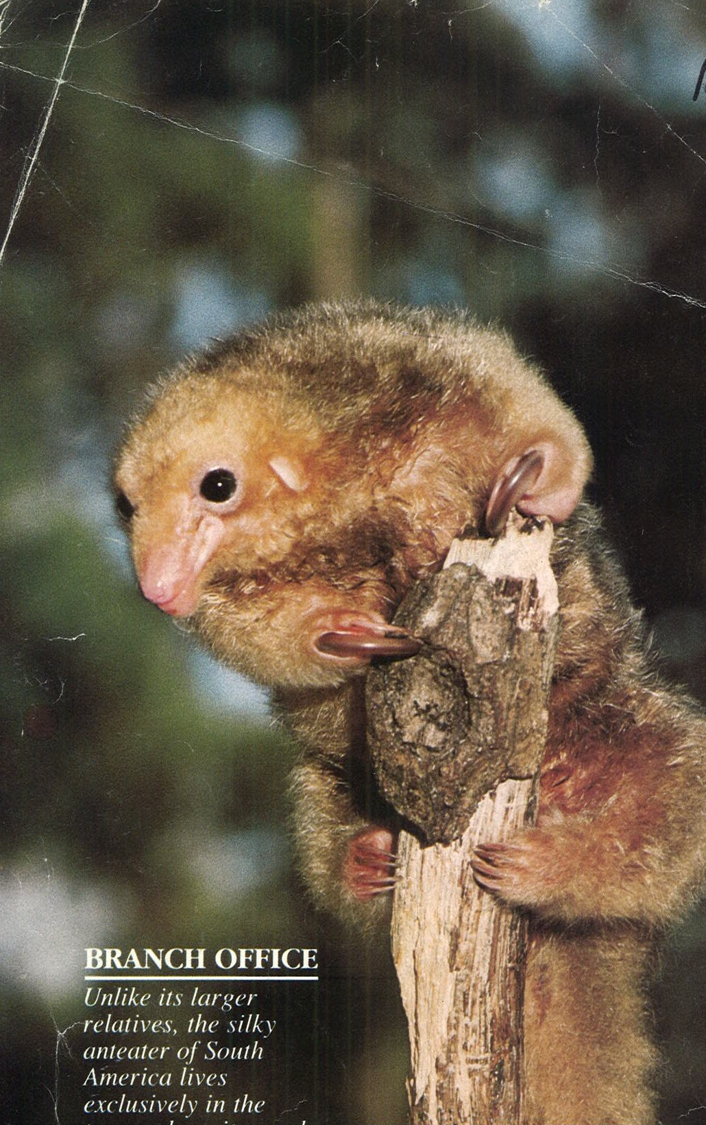
Anteater

National Geographic, September 2005.
Russian Desman

Albino Animals